The choice between a microscope and an endoscope depends on the disease, the ear’s anatomy and the work the surgeon must perform—not on which technique sounds newer. By the end, you will know what the two approaches involve, which test results influence the decision, what to ask at consultation and how to assess cost, risks and recovery.
Key takeaways
- The diagnosis, not the viewing method, determines whether ear surgery is needed.
- Ask how the surgeon will assess hearing, the eardrum, middle ear, and mastoid.
- Compare total treatment cost, follow-up visits, anaesthesia, and revision care.
- Discuss recovery time, hearing expectations, risks, and technique-specific limitations before consenting.
Microscopic and endoscopic ear surgery: what is the practical difference?
Microscopic and endoscopic ear surgery describe the viewing method, not a diagnosis or a promise of a smaller operation. The diagnosis must decide whether microscopic or endoscopic ear surgery in Bangalore is appropriate.
| Option | View and access | Practical strengths and limits |
|---|---|---|
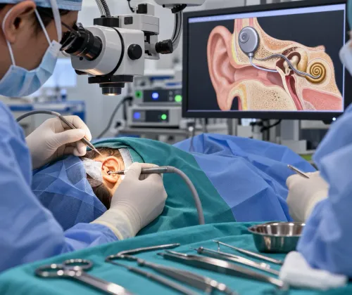
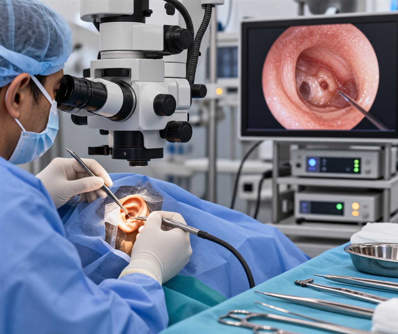
| Microscopic | A stable, magnified view from outside the ear canal | Leaves both hands free for drilling, grafting and reconstruction; supports prolonged drilling, extensive mastoid exposure and complex repair |
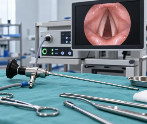
| Endoscopic | A wide-angle view from inside the ear canal | Shows recesses and areas hidden around corners in a straight microscopic view; the endoscope commonly occupies one hand, making prolonged drilling and complex reconstruction more demanding |
An endoscopic approach can avoid a postauricular incision in selected procedures. That does not make every endoscopic operation minor: it can still require drilling, grafting, ossicular reconstruction or mastoid work.
Microscopic surgery may offer better two-handed control when disease extends widely or reconstruction is intricate. Endoscopy may provide useful canal-based access and visibility, but a narrow canal, bleeding, swelling or disease beyond the useful line of sight can require microscopic or combined surgery.
Your surgeon should match the technique to the ear problem, anatomy and required repair—not choose it because one label sounds less invasive.
Which ear problems may need surgery, and which approach can treat them?
Surgery may be needed for chronic otitis media, a persistent tympanic-membrane perforation, cholesteatoma, retraction pockets, ossicular-chain damage, otosclerosis, selected ear-canal disease, or a middle-ear mass.
| Condition | Endoscopic approach | Microscopic or combined approach |
|---|---|---|
| Chronic infection or perforation | Disease reachable through the canal, with repair possible through that route | Extensive disease, poor access, or a need for mastoid exposure |
| Retraction pocket or cholesteatoma | Carefully selected disease visible and reachable around the canal | Disease extending beyond the useful line of sight or requiring wider clearance |
| Ossicular damage or otosclerosis | Selected reconstruction or stapes procedures | Complex reconstruction or limited instrument access |
| Ear-canal disease or middle-ear mass | Localised lesions accessible through the canal | Lesions needing wider exposure or control of hidden extension |
The same diagnosis can suit either technique in selected patients. In cholesteatoma, the priority is complete disease removal and a safe, dry ear—not whether the operation is labelled microscopic or endoscopic.
Endoscopic ear surgery in Bangalore may suit disease confined to areas reachable through the canal; microscopic or combined surgery may be safer when disease extends beyond the view or requires extensive mastoid exposure.
Observation, medical treatment, or hearing rehabilitation remains reasonable for some diagnoses. Ask which structure is diseased, what happens without surgery, and whether the proposed approach achieves infection control, a safe ear, hearing improvement, or all three.
How does the surgeon decide which technique fits your ear?
The surgeon chooses the technique after matching access to the diagnosis, not from the operation’s label.
| Approach | Strength | Limitation |
|---|---|---|
| Microscopic | Stable magnified view with both hands free for drilling, grafting and reconstruction | A straight view can hide recesses; a postauricular incision may be needed |
| Endoscopic | Wide-angle canal view that can reveal hidden recesses and avoid an external incision in selected cases | One hand holds the scope, making prolonged drilling or complex reconstruction harder |
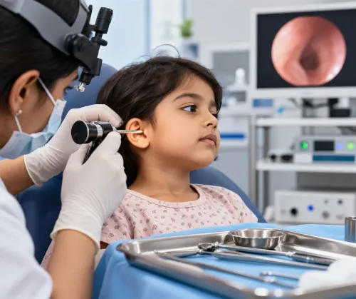
Otoscopy and ear endoscopy show the eardrum, canal, drainage, retraction and visible disease. Audiometry records air- and bone-conduction thresholds and speech understanding where appropriate; tympanometry assesses middle-ear function. A high-resolution CT of the temporal bone is reserved for suspected cholesteatoma, difficult anatomy, ossicular abnormality, complications, chronic disease or revision surgery.
It is not required for every uncomplicated perforation and cannot replace examination or audiology.
Use this checklist when choosing an ear surgery specialist in Bangalore:
- Exact diagnosis and hearing level
- Active infection and previous ear operations
- Canal anatomy and the surgeon’s otology or neurotology experience
- Whether grafting, ossicular reconstruction or mastoid surgery is needed
- Recognised ENT qualification and current medical registration
“Endoscopic” describes a viewing method, not proof of expertise in every ear operation.
What should happen during an ear surgery consultation in Bangalore?
A useful ear surgery consultation in bangalore starts with your symptoms, drainage, pain, hearing change, dizziness, tinnitus and every previous ear operation. The clinician examines the canal and eardrum, reviews audiometry, tympanometry and speech scores, and studies a high-resolution temporal-bone CT when disease, anatomy or revision surgery warrants it.
You should leave with a diagnosis, alternatives such as observation, medication or a hearing aid, and a specific goal: a safe dry ear, infection control, better hearing, or a combination.
| Approach | What it involves | When it may fit |
|---|---|---|
| Microscopic | External magnified view with two-handed access | Drilling, grafting, mastoid work or complex reconstruction |
| Endoscopic | Canal-based wide-angle view around recesses | Disease reachable through the canal |
| Combined | Uses both viewing methods | Disease needs wider access plus angled inspection |
Discuss local versus general anaesthesia, day-care or hospital admission, packing removal, water precautions, medication, cleaning, follow-up and whether a second-look or revision operation could be needed. Dr. Sohini's ENT Care - Empathy meets Expertise can provide the structured review needed before technique is selected.
- Why is surgery needed, and what happens without it?
- Is observation or a hearing aid reasonable?
- Which approach and why; will you use a graft or prosthesis?
- What are the facial-nerve, taste, dizziness, tinnitus and hearing risks?
- What does success mean, and when will audiometry be repeated?
Related services
OPD Consultation and Procedures Discover advanced and precise ear care with Dr. Sohini Chakrabarti's Ear Endoscopy services. Dr. View service → |
Surgical Procedures Ear Surgeries (Microscopic and Endoscopic) Embark on a journey to optimal hearing health with the expertise of Dr. View service → |
What are the limits, risks and recovery trade-offs of each approach?
Endoscopic surgery becomes a poor fit when a narrow or tortuous ear canal prevents instrument movement, bleeding or swelling obscures the lens, or one-handed access limits drilling and reconstruction. Extensive cholesteatoma, disease requiring wide mastoid exposure, or complex reconstruction can favour microscopic or combined surgery. An endoscope does not eliminate drilling, grafting or mastoid work.
Microscopic surgery provides a larger operative field and two-handed control, but it may require a postauricular incision behind the ear. That incision can add soreness, dressing care, wound-healing time and a longer recovery, even when it gives safer access to disease. Neither approach guarantees that disease will not persist or recur.
Possible outcomes include residual or worsened hearing loss, tinnitus, dizziness, infection, graft failure and revision surgery. Chorda tympani irritation can alter taste. Rare complications include facial-nerve injury and cerebrospinal-fluid leak. Repairing the eardrum does not guarantee normal hearing: the result also depends on the cochlea, ossicles and middle-ear ventilation.
Recovery remains active treatment. Expect packing removal, ear cleaning, prescribed topical treatment, water precautions and repeat audiometry. After cholesteatoma surgery, continued surveillance is essential because residual or recurrent disease can appear after the wound looks healed.
How should you compare cost and make the final decision?
There is no single clinically meaningful price for microscopic ear surgery cost in Bangalore. The estimate depends on the operation and the resources it requires.
| Operation | Cost drivers to check | Ask whether included |
|---|---|---|
| Tympanoplasty | Graft material, anaesthesia, hospital or day-care charges | Packing, medicines and follow-up |
| Mastoidectomy | Imaging, drilling, anaesthesia and hospital stay | Pathology and postoperative cleaning |
| Ossiculoplasty | Prosthetic implant, graft and audiology | Implant cost and later hearing tests |
| Stapes surgery | Prosthesis, anaesthesia and day-care charges | Revision-related fees |
| Cholesteatoma surgery | Imaging, pathology, mastoid work and surveillance | Second-look or revision surgery |
Request an itemised written estimate. Ask which charges apply if residual disease, graft failure or a planned second procedure makes revision necessary.
Do not compare published success percentages unless the endpoint is identical. A dry, safe ear; an intact graft; disease eradication; a target hearing threshold; and freedom from revision are different outcomes. Cholesteatoma removal and hearing restoration are separate goals.
Choose the approach that gives the surgeon adequate access to remove disease and complete reconstruction after reviewing examination, audiology, anatomy and imaging—not the approach marketed as scar-free or automatically safer.
Frequently asked questions
What is the practical difference between microscopic and endoscopic ear surgery?
Microscopic surgery uses a binocular operating microscope for depth perception and both-hand instrument work. Endoscopic surgery uses a slender camera through the ear canal, offering a wider angled view and access through smaller canal openings. The operation’s extent still depends on the diagnosis.
Which ear problems may need surgery?
Surgery can be considered for conditions such as persistent eardrum perforation, chronic middle-ear infection, cholesteatoma, troublesome ear discharge, conductive hearing loss from ossicle damage, or selected mastoid disease. Ear infections, wax, and many cases of hearing loss do not automatically require surgery.
What happens during an ear surgery consultation in Bangalore?
The consultation should include symptom history, ear examination, hearing tests such as pure-tone audiometry and tympanometry, and a review of imaging when needed. Ask about the diagnosis, proposed operation, technique, anaesthesia, risks, recovery, hearing expectations, follow-up, and total estimated cost.
How should you compare microscopic ear surgery cost in Bangalore?
Request an itemised estimate covering the surgeon, hospital, operating room, anaesthesia, medicines, hearing tests, imaging, follow-up visits, and possible revision treatment. Compare the proposed procedure and included care, not only the headline price.
Keywords